By delving into the innermost workings of cells, four MIT researchers who spoke at the technology track on life sciences and bioengineering at the ILP Research Directors Conference are delivering useful substances to cells to control disease, making progress on understanding how and why cells age, seeing living cartilage in the body to monitor the progression and treatment of degenerative disease, and uncovering the secret of cancer cells' mind-boggling ability to reproduce themselves forever.
CONFERRING IMMORTALITY
Robert A. Weinberg, the Ludwig Professor for Cancer Research and the American Cancer Society Professor of Biology, described how cancer cells -- unlike normal cells -- need no influx of growth factors to multiply indefinitely in a petri dish. They make their own growth factors, which they release outside the cell and which in turn stimulate the cell to produce more growth factor.
There must be at least two changes to a cell to transform it from a normal cell to a cancer cell, said Professor Weinberg, a founding member of the Whitehead Institute for Biomedical Research. It must develop the ability to produce its own growth factor, and it must learn to resist the inhibitory signals from neighboring cells that keep normal cells from proliferating out of control.
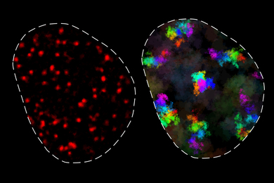
The third insidious characteristic of cancer cells is that they do not have the "generational clock" of normal cells that stop them from growing after a certain number of generations. While normal cells seem programmed to stop multiplying after 50 or 60 generations, cancer cells, with enough space and nutrients, will simply grow forever. This raised the question of how normal cells "know" how many times they had doubled in the past.
The answer seems to lie within the ends of chromosomes, called telomeres. These tips, made up of specific gene sequences, protect the chromosomes from damage like plastic tips protect the ends of shoelaces, Professor Weinberg explained.
When cells replicate, they fail to copy the entire telomere sequence, so with each reproduction, the telomeres grow a little shorter. This leads to cell death. Scientists speculate that this innate ability of cells to bring about their own demise is a protective mechanism that keeps premalignant cell populations from turning cancerous.
However, about one cell in a million solves the problem of telomere collapse by generating an enzyme called telomerase. Cancer cells are the only cells that produce this enzyme, effectively creating their own means to immortality.
Professor Weinberg's recent efforts to "immortalize" normal cells by giving them the ability to make telomerase led to cells that grew for a far longer time than normal cells, but they did not proliferate indefinitely. "We believe we may learn how to immortalize these cells," he said, leading to a better understanding of the genetic basis of human cancer.
'SEEING' INSIDE LIVING TISSUE
Martha L. Gray, the J.W. Kieckhefer Associate Professor of Electrical Engineering and co-director of the Harvard-MIT Division of Health Science and Technology, described her team's progress on watching cartilage degradation and repair. To Professor Gray and others trying to "see" the 1 to 2mm of sponge-like tissue called cartilage that encases our bones, an X-ray is of little help. Cartilage damage might show up as a decreased space between two bones, but that's not much to go on when you're trying to diagnose and treat a case of arthritis so severe that the tissue has been eroded to nothing.
With magnetic resonance imaging (MRI), however, one can see cartilage and look for the absence or disruption of tissue. Taking that a step further, Professor Gray has come up with a way to measure the concentration of ions within the fluid of affected tissue. The ions indicate the presence or absence of proteoglycans -- brush-shaped molecules that give cartilage some of its bone-protective qualities.
By using MRI data to quantify the concentration of ions, researchers have found a way to "see" living cartilage, detect any changes before the tissue is badly disrupted, monitor disease progression and choose appropriate therapeutic strategies.
AGING CELLS
"What is it like to be an old cell?" asked Professor of Biology Leonard P. Guarente, who addressed the molecular cause of aging. He is especially interested in old yeast. While most of us can't tell young yeast from old yeast, researchers can do so by watching the cells divide. They divide by budding, with a daughter cell of new material breaking off from the mother cell, which gets older with every division. The mother cell can divide 20 times before it stops.
By watching yeast divide, Professor Guarente has found that there are changes in the nucleolus -- a section of the nucleus -- of cells that are aging. He has found that in the nucleolus of older cells, some of the cell's genetic material, a circular piece of ribosomal DNA, pinches off from a chromosome and accumulates in the cell, causing it to enlarge.
These "circles" of material happen with aging and at the same time cause aging, he said. The circles double with every cell division, growing exponentially, producing more and more ribosomal proteins that poison the cell and eventually kill it.
They solve a short-term problem for the cell -- trying to repair damage to rDNA -- while establishing its mortality. One of the next steps is to determine if the same process happens in human cells that, like yeast, undergo asymmetrical cell division. These kinds of cells are found in the skin, kidney and liver as well as the blood.
AN ENGINEERING APPROACH
Douglas A. Lauffenberger, the Joseph R. Mares Professor of Chemical Engineering and director of the Center for Biomedical Engineering, said an engineering approach to manipulating cell function can provide a more effective way to deliver useful substances to cells.
Once a useful substance like a growth factor or a protein binds with a cell, many dynamics can occur, he said. Researchers in biomedical engineering consider the interaction of all the components of the cell, like the many components of an engineering system. "If you can control one step, you can do as much or more at a systemic level," he said.
A version of this article appeared in MIT Tech Talk on May 13, 1998.